Introduction: Enhancing Brain Tumor Image Classification Using Deep Learning and Image Augmentation Techniques
 Mehdi Mehri
Table of contents

This 4 parts series of articles excluding the introduction, will go through the development of my proposed system for cerebral tumor classification which I worked on for my master's degree.
For my final year, I wanted to work on a big project that would be a showcase of all the things I learned up until this year and wanted to keep learning about in the future. Finding a subject to work on that would be challenging and put me out of my comfort zone yet still manageable proved to be a rather complicated task.
After doing some research about different types of data that I wanted to work on I finally decided to get my hands dirty for the first time with cerebral MRI data. Up until that point, I had little experience with image data with just a handful of projects related to recognition and segmentation. So I just had enough experience to know for sure that I'd enjoy working with medical imaging but I wasn't ready for the challenges that I was about to face in the upcoming months.
Prerequisite Knowledge
In order to understand the concepts discussed in this series, you'll need to have some basic understanding of the following:
Image data manipulation and processing.
Image data augmentation techniques such as geometric transformations and generative augmentation.
Feature extraction and transfer learning.
Machine learning models creation and training.
1. Structure of the Series
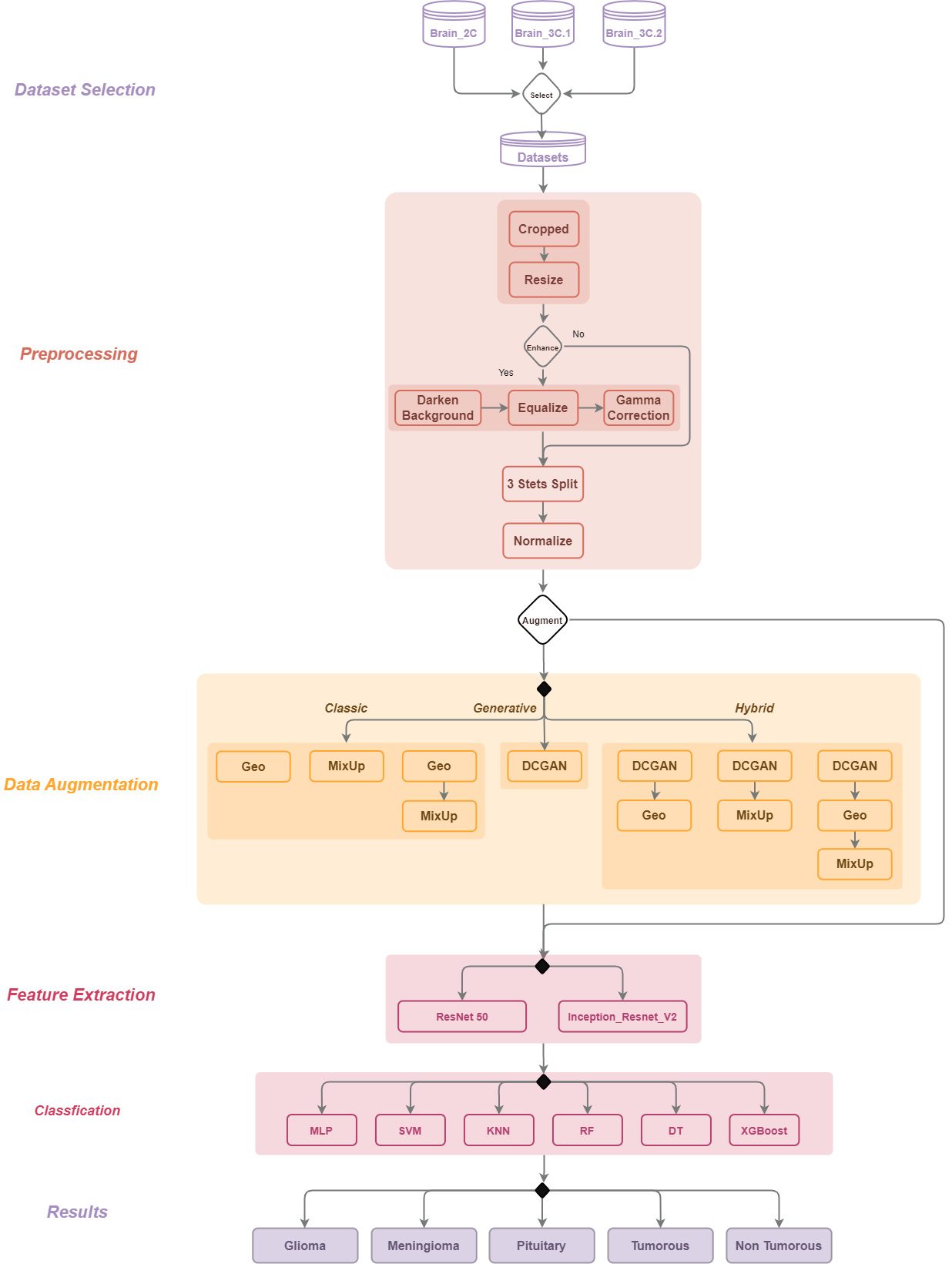
Each part of the series will be dedicated to one section of the system architecture:
1.1. Image Data Preprocessing
This is an essential part of any deep-learning system that deals with images, it manipulates the image and simplifies them to facilitate the job of the following steps.
1.2. Image Data Augmentation
This step will increase the data that will be passed to the following steps in order to improve the performance of the model training.
1.3. Feature Extraction
Once the images are processed we pass them to a deep-learning feature extractor. For this system, we opted for a transfer learning approach using DL models pre-trained on a novel MRI dataset called RadImageNet (Reference).
1.4. Classification
Finally, we apply popular classifiers such as MLP, KNN XGBoost, to classify the images into one of the five studied classes: Glioma Meningioma, Pituitary, Tumorous, and Non-Tumorous.

General System Architecture
2. Introduction to the datasets
This study uses 3 datasets to do tests and experiments on the proposed architecture in 3 different augmentation and classification conditions.
Note that since the datasets used do not have agreed upon names or labels in the research community, we will be referring to them as Brain_2C, Brain_3C.1, and Brain_3C.2, with the C symbolizing the class number in the dataset.
2.1. Brain_2C (Balanced Binary)
The third dataset utilized in this study is sourced from Kaggle and includes a balanced set of medical images. The dataset is divided into two classes: tumorous and non-tumorous. Each class has a total of 1500 samples. The images in this dataset are all in the axial plane.
| Tumor Type | Number of Samples |
| Tumorous | 1500 |
| Non-Tumorous | 1500 |
| Total | 3000 |
2.2. Brain_3C.1 (Balanced Multi-class)
The second dataset considered in this study was sourced from Kaggle. We modified it to turn into a balanced dataset by removing the “non_tumorous” class.
The dataset includes three different tumor types: glioma, meningioma, and pituitary tumors, in addition to a fourth class for non-tumorous scans, with 926 samples, 937 samples, 901 samples, and 500 respectively. The total size of the dataset is 2764 samples. In addition to the balanced tumor samples, the dataset includes an additional class of non-tumorous brain scans that are not utilized in this research.
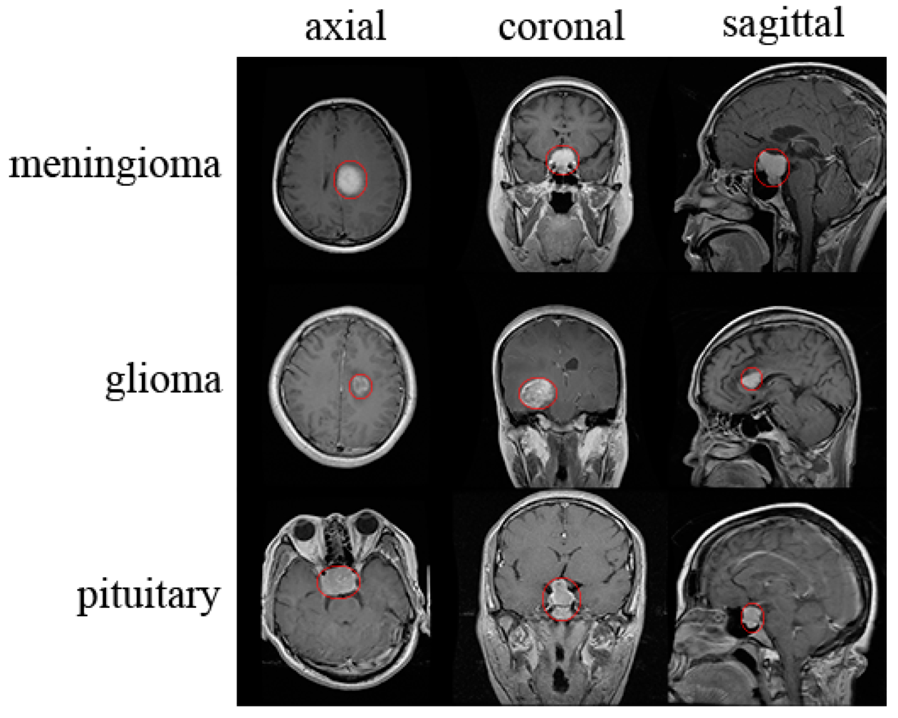
The images in the dataset are taken from multiple planes. The balanced nature of this dataset makes it an excellent resource for developing and testing models in stable and balanced conditions.
| Tumor Type | Number of samples |
| Glioma | 926 |
| Meningioma | 937 |
| Pituitary | 901 |
| Non-Tumorous | 500 |
| Total | 3264 |
2.3. Brain 3C.2 (Imbalanced Multi-class)
One of the datasets under consideration in this study was obtained from Jun Cheng's work on tumor segmentation research in 2015 “Brain Tumor Dataset”.
It comprises a total of 3064 T1-weighted contrast-enhanced images that were sourced from 233 patients' tumors available in 3 planes (axial, sagittal, and coronal). The dataset includes three different tumor types: glioma, meningioma, and pituitary tumors. The glioma class contains 46.5% of the total samples (1426), the meningioma class has 23.1% of the total samples (708), and the pituitary class contains 30.4% of the total samples (930). The class distribution of the dataset is highly imbalanced, with a ratio of 2.14:4.32:3.29 for glioma, meningioma, and pituitary, respectively. The dataset is composed of images taken from multiple planes.
This dataset provides an interesting challenge to showcase the capabilities of the models we used along with augmentation techniques.
| Tumor Type | Number of samples |
| Glioma | 708 |
| Meningioma | 1426 |
| Pituitary | 930 |
| Total | 3064 |
Conclusion
If all of the above sounds interesting then you're in for a treat, I'll be releasing Part I this week with the other parts next week. I hope you find these articles valuable and if you have any questions don't hesitate to comment, I'll do my best to answer you in detail.
Subscribe to my newsletter
Read articles from Mehdi Mehri directly inside your inbox. Subscribe to the newsletter, and don't miss out.
Written by

Mehdi Mehri
Mehdi Mehri
I'm a passionate computer science student with a strong academic background in Artificial Intelligence and Data Processing. Holding a Master's degree in AI with excellent grades, I've dedicated my academic journey to exploring the fascinating world of AI and its applications. 🧠 During my final year, I delved deep into the intersection of AI and healthcare, particularly in medical image analysis. My research project, titled "Enhancing Brain Tumor Image Classification Using Deep Learning and Image Augmentation Techniques," reflects my commitment to solving real-world challenges with cutting-edge technology. 🔍 This research not only earned me academic recognition but also led to the creation of a manuscript that I aim to publish in a scientific journal. I'm excited to contribute to the field of AI and healthcare, bridging the gap between technology and improving patient outcomes. 🤝 Currently, I'm actively seeking internship opportunities in Data Science and Artificial Intelligence, where I can apply my knowledge and skills to real-world projects. If you're interested in collaborating or discussing AI-related opportunities, feel free to connect with me!
