What I learned building a healthcare product
 Prashanth Shenoy
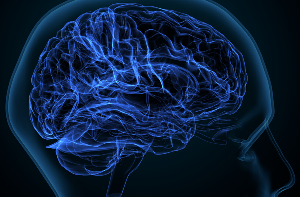
(Picture courtesy Penn Medicine)
Building a healthcare product at times leads an Engineer to a fascinating convergence of Physics, Biology and Technology. How far you go down the rabbit hole of curiosity depends on your interest and the time you are willing to invest in it.
In this blog post I would like to share some of my learnings building an EEG diagnostic solution. An EEG system is used for diagnosing brain tumor, sleep disorders, dementia and many other conditions.
An EEG (Electroencephalogram) system measures the electrical activity in the brain. A person undergoing EEG scan has electrodes attached to their scalp.
(Picture credit Mayo Clinic).
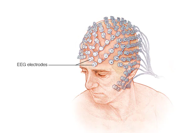
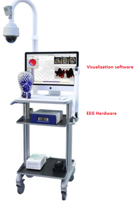
The electrodes measure the electrical activity of brain cells. These electrical signals are then amplified by hardware attached to the electrodes. The electrical signals are then visualized using software.
This is what an EEG system looks like.. (Picture courtesy EGI)
Each brain cell (neuron) is a tiny battery
If I recollect well, in high school we were taught brain's anatomy and its modular structure. We were told that different parts of the brain are responsible for distinct functions such as memory, movement and vision. However the electrical properties of the brain were either glossed over or perhaps I was not paying attention.
Here are some examples of EEG signals that you would see in the "visualization software" mentioned above.
- EEG waveforms captured when a person in a "resting state" with eyes closed.
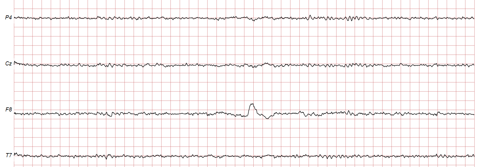
EEG waveforms captured when a person is listening to music in a native language for 3 minutes with headphones on.
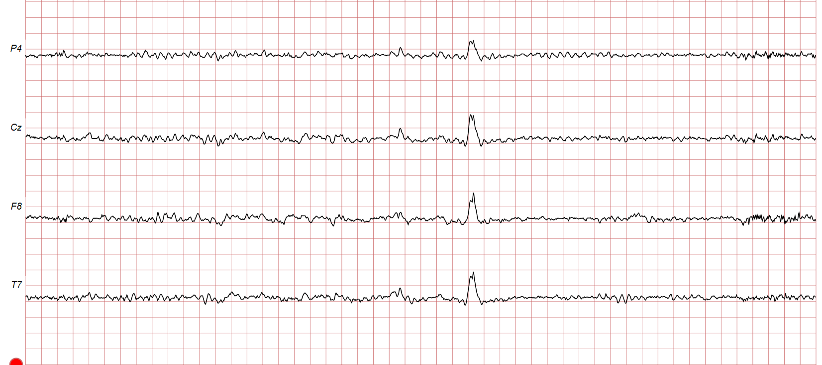
EEG waveforms captured when a person is listening to music in a non-native language for 3 minutes with headphones on.
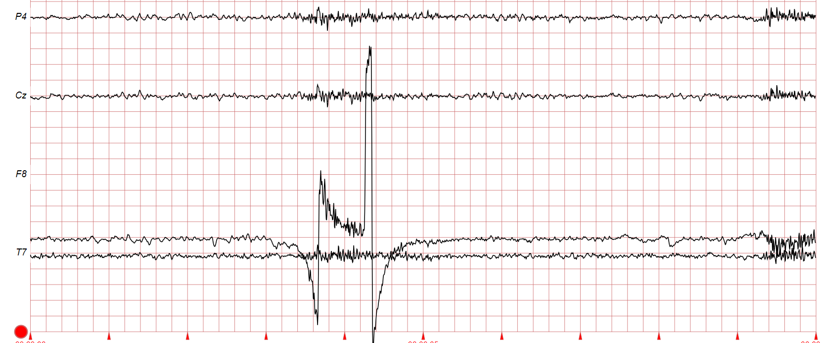
(The images are screenshots of data that can be found here ->Physionet)
Even without any sort of medical background, one can easily deduce that the EEG signals reveal something about the mental state of a person.
The realization that neurons are a bag of chemicals, the movement of simple sodium and potassium ions across cell walls and Neurotransmitters across a synapse , causes a change in electric potential and gives rise to intensely personal emotions such as happiness, anxiety, attention, boredom made me contemplate on the nature of mind. Its fascinating and humbling at the same time.
Brain chooses to represent body differently.
We have a picture of our body in our mind and this gets reinforced whenever we look at ourselves in the mirror. When I started reading more about brain I came across this wonderful book called Phantoms in the Brain by VS Ramachandran. I learned that how the brain chooses to represent our body is completely different.
If mild electrical stimulation is applied to different parts of the outermost layer of the brain (called the cerebral cortex) a corresponding body part starts moving/twitching/experiencing sensation. Crudely put this is like reverse EEG, where instead of measuring electric potential of neurons we are stimulating the brain electrically.
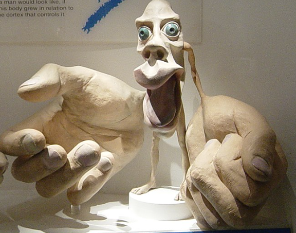
If one were to visually represent the human body in proportion to the amount of brain tissue dedicated to it for processing sensory function, we end up with something called Penfield Homonculus. (Picture courtesy Wikipedia).
Notice how large the hands are represented in the brain. This disproportionate representation is because of the fine motor control that our hands require.
Human brain is complex yet fragile (sometimes)
The British neurologist Oliver Sacks wrote the famous book The Man Who Mistook His Wife for a Hat . This book through a collection of clinical case studies describes how the concepts of human identity, brain anatomy, and perception of time are all deeply intertwined to make us who we are.
One case in the book describes a patient with Visual Agnosia, a condition where the individual can see objects but cannot recognize them. For example, the patient might see a painting of a serene natural landscape and see only abstract geometric shapes, without ever recognizing trees, rivers or other elements.
One of the causes of Visual Agnosia is stroke. A stroke is a disruption in blood supply to the brain. When the brain is deprived of oxygen the brain cells begin to die.
The product that we were building was for long term epilepsy monitoring. Epilepsy is a disorder caused by abnormal nerve cell signaling, leading to seizures. Seizures are uncontrolled bursts of electrical activity in the brain that can affect sensations, behaviors, awareness, and muscle movements.
Impact of technology
I do not have an iota of background in medicine, however I was fascinated by images such as the following. (Such images were part of the dataset shared by our product manager for product QA)
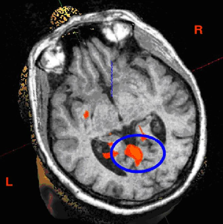
The above image is an fMRI image. (Picture courtesy https://www.intechopen.com/chapters/45187).
fMRI is used to help identify the areas of the brain that are responsible for generating seizures. By identifying this zone, doctors can better plan surgical interventions to remove or treat the area causing the seizures. fMRI is also used in assessment and management of stroke (described in the previous section)
The physics behind fMRI is deep and there is a lot of engineering heft behind it. The machines cost north of a million dollars. The essence of fMRI is to measure brain activity by detecting changes in blood flow. Here is a great video explaining the basics of fMRI.
Role of serendipity in learning
This was a time before LLMs burst into the world. I was searching for information online to understand more about fMRI and ended up discovering the brilliant Nancy Kanwisher. How I wish I had a teacher like this!
The essence of learning I think is in engaging with lectures such as this, with a sense of scientific curiosity, humbly recognizing that your understanding is a mere droplet in the ocean of knowledge. It's a privilege to live in an era where such valuable content is freely accessible to us!
Subscribe to my newsletter
Read articles from Prashanth Shenoy directly inside your inbox. Subscribe to the newsletter, and don't miss out.
Written by

Prashanth Shenoy
Prashanth Shenoy
I have professionally contributed to the following products: https://www.hsdp.io/discover/2net https://www.philips.ae/healthcare/resources/landing/tasy https://www.philips.com.qa/healthcare/product/HC1402561/geodesic-eeg-system-400-research-high-density-eeg-system https://www.philips.com/c-dam/b2bhc/master/Products/Category/enterprise-telehealth/mobile-obstetrics-monitoring/452299112911_MOM_WhitePaper_HR.pdf https://www.philips.co.in/healthcare/innovation/about-health-suite https://www.philips.co.in/healthcare/solutions/radiation-oncology/radiation-treatment-planning