Article Review: New compartment model for hepatic blood flow quantification in humans from 15O water PET images
 Aldo Yang
3 min read
Objectives
- Introduces a new dual-input compartment model for hepatic blood flow (HBF) quantification using 15O-water PET images.
- Utilizes TotalSegmentator for automatic segmentation of aorta, hepatic portal vein (PV), liver, and spleen to obtain image-derived input functions (IDIFs).
- Compares the new model with three existing models: one-tissue compartment model (1TCM) and two versions of the dual-input model.
- Shows that the new model performs best in terms of mean relative error (MRE) (p-values≤0.001).
- Estimates arterial, portal, and total HBF, finding moderate positive correlation between arterial and portal HBFs and negative correlation between total HBF and patient weight.
Methodology
- Automatic segmentation of regions of interest (aorta, hepatic PV, liver, spleen) using TotalSegmentator, a deep learning-based tool, on CT images.
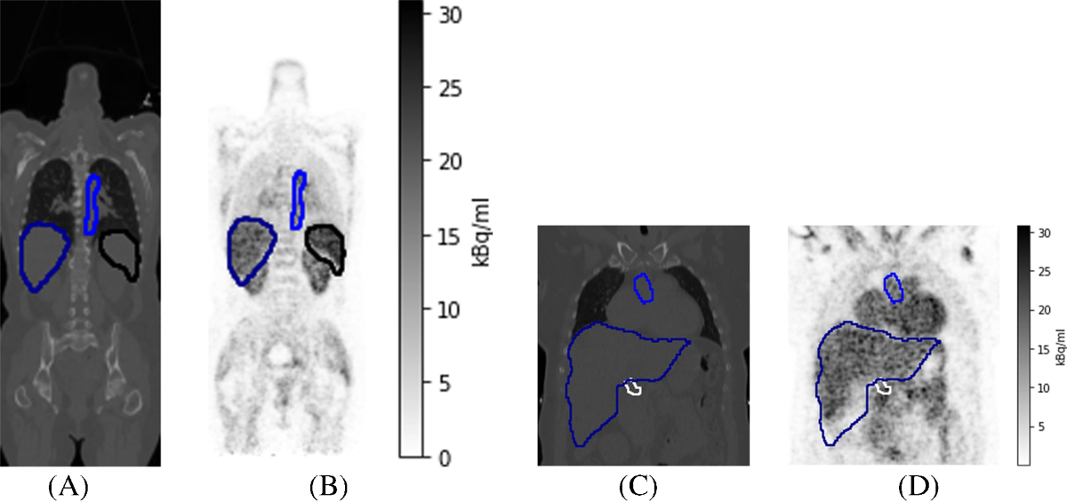
- Extraction of mean time-activity curves (TACs) from the segmented regions.
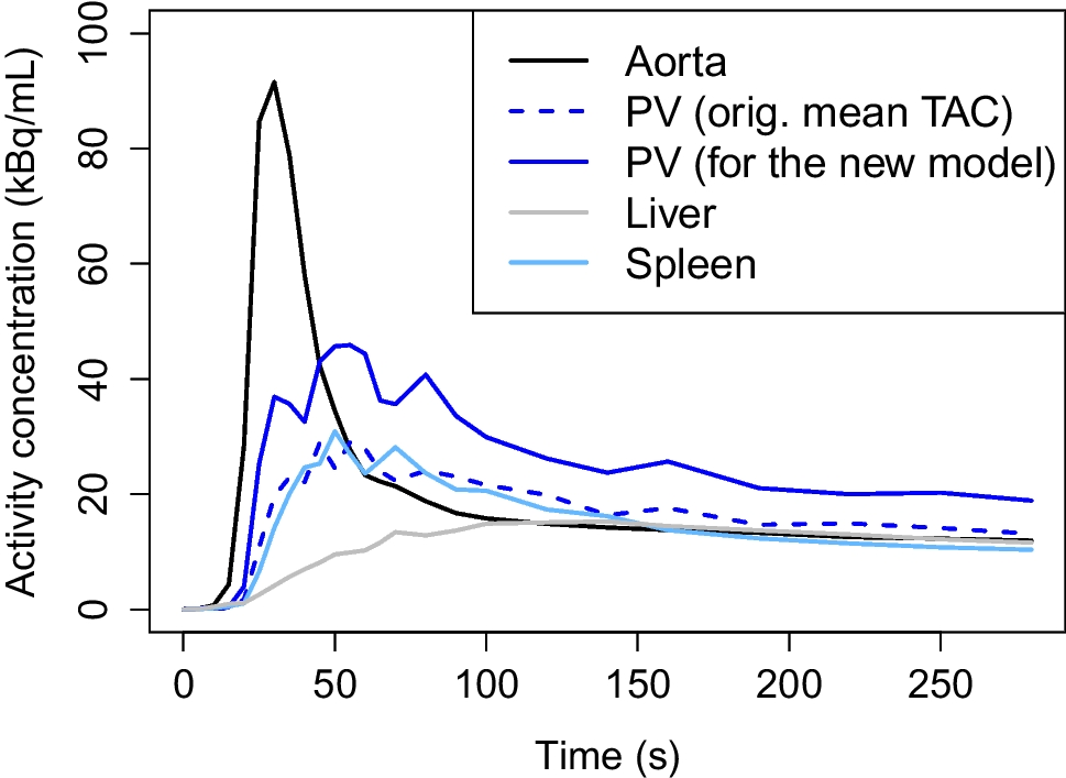
- Modification of the hepatic PV TAC by using only voxels above the 90th percentile to mitigate respiratory motion artifacts and potential underestimation.
- Development of a new dual-input compartment model with IDIFs from aorta and hepatic PV.
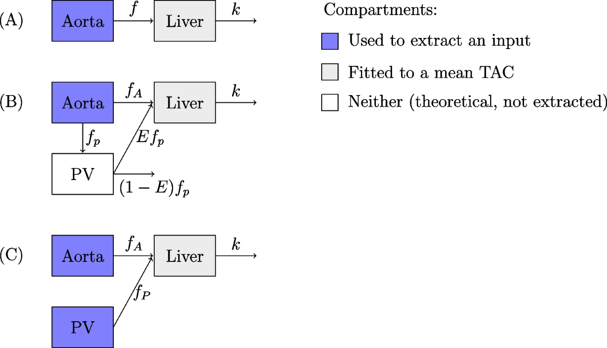
- Comparison of the new model with 1TCM and two existing dual-input models (Taniguchi et al. and Rijzewijk et al.).
- Model fitting using a non-linear Newton-type minimization algorithm (nlm in R) to minimize the sum of squared differences between the measured and model-predicted liver TACs.
- Use of absolute value and inverse-logit functions to constrain parameter ranges.
- Selection of time delay parameters based on the lowest model error.
Results
- The study used 15O-water PET data from 57 patients.
- The new model provided mean arterial HBF of 0.299 ± 0.168 mL/min/mL, mean portal HBF of 0.930 ± 0.520 mL/min/mL, and total HBF of 1.229 ± 0.612 mL/min/mL.
- The new model's performance was superior to the other models in terms of MRE (p-values ≤ 0.001).
- The ratio of portal HBF to total HBF (74.3 ± 11.5%) was consistent with literature values (e.g., 75% reported by Sureka et al.).
- Moderate positive correlation (0.438, p-value ≤ 0.001) was found between arterial and portal HBFs.
- Moderate negative correlation was found between total HBF and weight (-0.431, p-value ≤ 0.01) and BMI (-0.351, p-value ≤ 0.01).
- No significant sex- or age-based differences were found in HBF.
Discussions
- The study's reliance on TotalSegmentator for PV segmentation is a potential limitation. The small size of the PV, even with the improved resolution of the Biograph Vision Quadra, makes accurate segmentation challenging. Further validation of the PV segmentation against manual segmentation or other imaging modalities (e.g., MRI) would strengthen the findings.
- The 90th percentile threshold for PV TAC calculation, while intended to address motion artifacts, introduces a potential bias. Investigating the sensitivity of the results to this threshold and exploring alternative motion correction techniques would be beneficial.
- The study acknowledges the potential for overestimation of HBF due to underestimation of PV concentration. Exploring partial volume correction methods could improve the accuracy of the HBF estimates.
- The study population consisted of patients with suspected coronary artery disease, which may limit the generalizability of the findings to healthy individuals. A comparison with a healthy control group would be valuable.
- While the study found a negative correlation between HBF and weight/BMI, the underlying mechanisms are not fully elucidated. Further investigation into the potential confounding effects of comorbidities (e.g., type 2 diabetes) is warranted.
Reference: New compartment model for hepatic blood flow quantification in humans from 15O water PET images
0
Subscribe to my newsletter
Read articles from Aldo Yang directly inside your inbox. Subscribe to the newsletter, and don't miss out.
Written by